Track your pregnancy journey with at-home fetal Dopplers. Analyze ultrasound biophysics, gel coupling, and clinical safety guidelines.
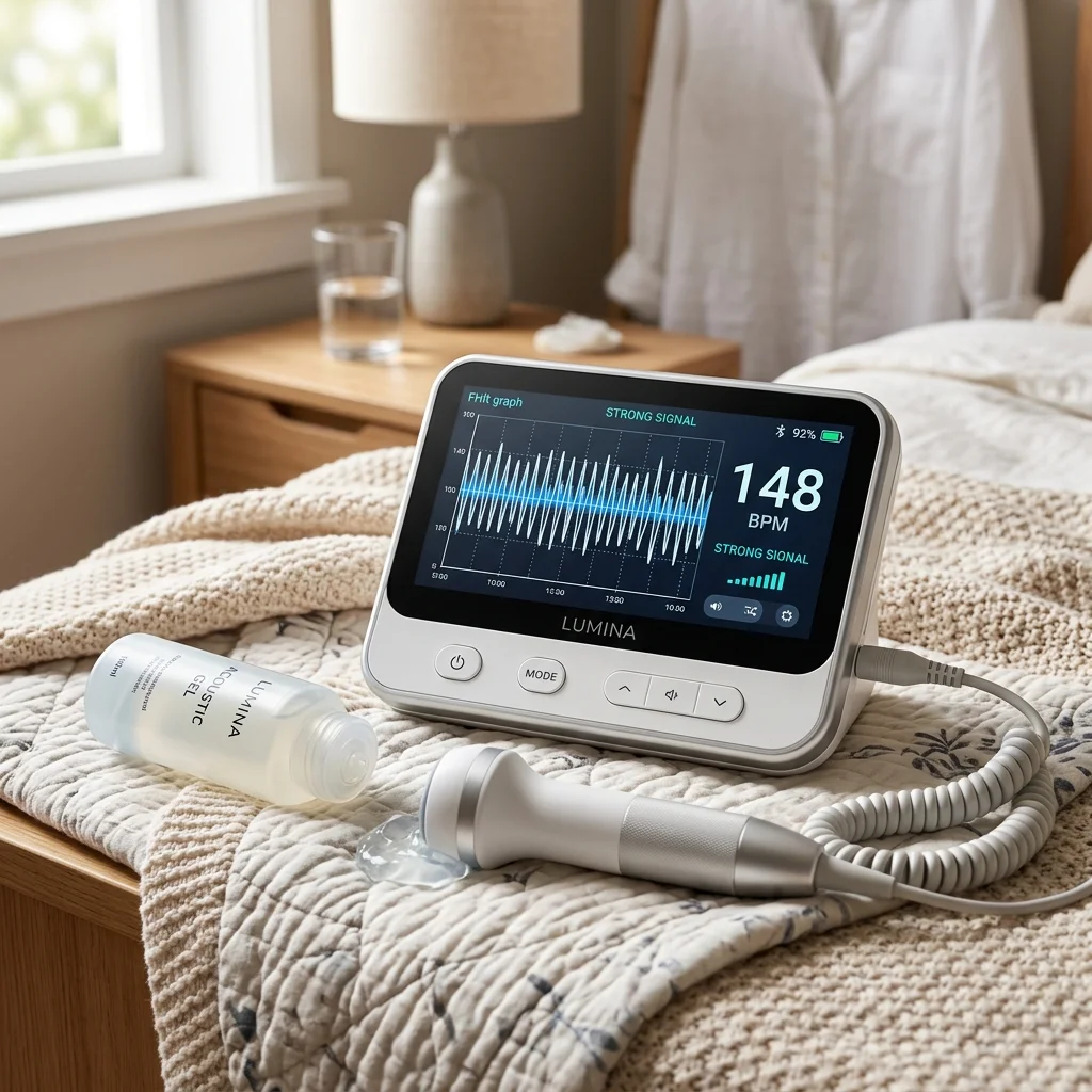
- High-sensitivity 3MHz probe delivers clear fetal heart sound detection
- Backlit LCD display shows real-time fetal Heart Rate (FHR) calculations
- Built-in speaker with volume control and audio output jack for headphones
Clinical & Performance Insights
Track your pregnancy journey with at-home fetal Dopplers. Analyze ultrasound biophysics, gel coupling, and clinical safety guidelines.
1. Physics of Ultrasound and the Doppler Effect
At-home fetal Dopplers rely on the physics of ultrasound waves. The device's probe contains piezoelectric crystals that convert electrical energy into high-frequency sound waves. These waves travel through the abdomen and reflect off internal structures.
The Doppler effect occurs when sound waves reflect off a moving object, causing a frequency shift. In pregnancy monitoring, the moving target is the beating fetal heart and closing valves. The probe detects the reflected, shifted waves and converts them back into signals.
The monitor's processor analyzes these signals to generate an audible heartbeat sound and calculate heart rate. A 2 MHz to 3 MHz probe is typically used to balance tissue penetration and resolution. Understanding this physics helps users operate the device correctly.
Ultrasound wave frequency determines the resolution and penetration of the signal. A 3 MHz probe is optimized for early pregnancy, providing clear heart sounds. Understanding the biophysics supports accurate monitoring.
A 3 MHz probe provides high resolution for detecting heart beats in early pregnancy. The probe emits low-power waves that reflect off moving heart valves. Biophysical principles support accurate monitoring.
A 3 MHz probe provides high resolution for detecting heart beats in early pregnancy. The probe emits low-power waves that reflect off moving heart valves. Biophysical principles support accurate monitoring.
- Piezoelectric crystals convert electrical energy into high-frequency ultrasound waves.
- The Doppler effect shifts sound wave frequencies when reflecting off the beating heart.
- Reflected waves convert to electrical signals, generating an audible heartbeat sound.
- A 3 MHz probe balances penetration depth with signal resolution for home use.
- Understanding ultrasound physics helps users operate the Doppler probe effectively.
2. Fetal Heart Rate (FHR) Frequency Detection
A healthy fetal heart rate (FHR) typically ranges from 110 to 160 beats per minute. This rate is significantly faster than the maternal heart rate, which ranges from 60 to 100 BPM. The FHR sound has a rapid, galloping rhythm.
Urinary or placental blood flow can create a swishing sound that can be mistaken for the heartbeat. The maternal pulse can also be picked up, leading to false reassurance. Distinguishing these sounds requires practice and careful listening.
Modern Dopplers feature LCD screens that calculate and display the heart rate automatically. This calculation updates in real-time, helping confirm that you have located the fetal heart. Knowing target heart rate ranges supports accurate home monitoring.
Distinguishing fetal heart sounds from placental blood flow requires practice. The fetal heartbeat is fast and rhythmic, while blood flow creates a swishing sound. LCD displays help confirm the heart rate.
Distinguishing fetal heart sounds from placental blood flow requires practice. The fetal heartbeat is fast and rhythmic, while blood flow creates a swishing sound. LCD screens display calculations to help confirm.
Distinguishing fetal heart sounds from placental blood flow requires practice. The fetal heartbeat is fast and rhythmic, while blood flow creates a swishing sound. LCD screens display calculations to help confirm.
- A normal fetal heart rate ranges from 110 to 160 BPM, resembling a horse gallop.
- Maternal pulse is slower (60-100 BPM) and matches the mother's wrist pulse.
- Blood flow in the placenta can create a swishing sound that mimics a heartbeat.
- LCD displays calculate and show the fetal heart rate in real-time.
- Confirming the heart rate range prevents confusing maternal and fetal signals.
3. Acoustic Impedance and Gel Coupling
Ultrasound waves travel poorly through air, which has high acoustic impedance. When the probe is placed on dry skin, almost all ultrasound energy is reflected at the air-skin boundary. This reflection prevents the waves from penetrating tissues.
To solve this, a gel is used to match acoustic impedance between the probe and skin. The gel eliminates air gaps, allowing ultrasound waves to pass into the abdomen. Using gel is required to get a clear, audible signal.
Water-based gels are preferred because they do not stain clothing or damage the probe. Avoid using oils or lotions, which can degrade the probe's silicone seal and reduce signal quality. Proper gel application is crucial for successful monitoring.
Applying sufficient acoustic gel is the most important step for clear signals. The gel eliminates air gaps, letting ultrasound waves enter the abdomen. Water-based gel protects the probe and skin.
The coupling gel matches acoustic impedance, letting waves pass into the abdomen. Using gel is required to get a clear, static-free heart sound. Water-based gel protects the probe's silicone seal.
The coupling gel matches acoustic impedance, letting waves pass into the abdomen. Using gel is required to get a clear, static-free heart sound. Water-based gel protects the probe's silicone seal.
- Air has high acoustic impedance, reflecting ultrasound waves at the skin surface.
- Acoustic gel matches impedance, eliminating air gaps to let waves pass.
- Water-based gels are required to get a clear, static-free heart sound.
- Avoid lotions or oils, which can degrade the probe's protective silicone seals.
- Applying sufficient gel is the most important step for successful signal tracking.
4. Diagnostic vs. Screening Ultrasound Safety
Ultrasound is non-ionizing radiation, making it safer than X-rays. However, ultrasound waves transfer energy to tissues, which can cause slight heating. This thermal effect is measured by the thermal index (TI) on medical devices.
At-home Dopplers operate at low power levels to minimize thermal risk. Still, regulatory bodies recommend limiting use to prevent unnecessary tissue exposure. Keep sessions short, typically under 5 minutes, and avoid daily use.
Using the device as a screening tool, not a diagnostic one, supports safe home monitoring. It should supplement, not replace, regular prenatal visits with your obstetrician. Understanding safety guidelines helps maintain a healthy pregnancy journey.
FDA-cleared Dopplers operate within safe power limits to prevent tissue heating. Limit home monitoring sessions to a few minutes to ensure safety. Following guidelines protects your pregnancy journey.
Ultrasound transfers thermal energy to tissues, requiring prudent use of Dopplers. Keep home monitoring sessions under 5 minutes to ensure safety. Following guidelines protects mother and baby.
Ultrasound transfers thermal energy to tissues, requiring prudent use of Dopplers. Keep home monitoring sessions under 5 minutes to ensure safety. Following guidelines protects mother and baby.
- Ultrasound is non-ionizing radiation but can transfer thermal energy to tissues.
- FDA-cleared Dopplers operate at low power to prevent tissue heating risks.
- Limit home sessions to 5 minutes to avoid unnecessary ultrasound exposure.
- Dopplers are screening tools that supplement, not replace, obstetrician checkups.
- Following clinical safety guidelines ensures a healthy, low-risk pregnancy.
5. Home Monitoring and Anxiety Management
Home Doppler monitoring can provide reassurance, but it can also cause anxiety if the heartbeat cannot be located. Factors like fetal position, placenta placement, or maternal tissue thickness can block signals. Failing to find the heartbeat is common, especially in early pregnancy.
Users may feel anxious or make unnecessary emergency calls if they cannot locate the heartbeat. It is important to stay calm and try again later if the signal is elusive. The device is not a replacement for professional clinical evaluations.
Educating yourself on the limitations of home monitoring helps manage expectations. If you experience decreased fetal movement or other warning signs, contact your healthcare provider immediately. Reassurance should balance with clinical awareness.
Stay calm and try again later if the fetal heartbeat is difficult to locate. Uterine position and fetal movement can block the ultrasound signal. The Doppler is a reassuring, supportive tool.
Fetal movement and placenta placement can temporarily block the Doppler signal. Failing to locate the heart beat is common and does not indicate an emergency. Stay calm and try again later.
Fetal movement and placenta placement can temporarily block the Doppler signal. Failing to locate the heart beat is common and does not indicate an emergency. Stay calm and try again later.
- Fetal position, tissue thickness, and placenta placement can block signals.
- Failing to locate the heartbeat is common and does not indicate an emergency.
- Stay calm and try again later if the heartbeat signal is difficult to find.
- Do not use the Doppler as a substitute for professional diagnostic medical visits.
- Managing expectations reduces stress, supporting emotional health during pregnancy.
6. Identifying Fetal Distress Indicators
A persistently high or low fetal heart rate can indicate fetal distress. Tachycardia is an FHR over 160 BPM, while bradycardia is an FHR under 110 BPM. These conditions require immediate professional evaluation.
However, temporary variations in heart rate are normal and indicate fetal activity. The heart rate may speed up during movement and slow down during rest. Recognizing these normal patterns prevents unnecessary worry during monitoring.
Do not rely solely on the home Doppler to diagnose fetal distress. If you suspect an issue or notice changes in fetal movement, seek medical attention. The Doppler is a supportive tool, not a diagnostic device.
Recognizing normal heart rate fluctuations during fetal movement prevents unnecessary worry. The heart rate normally speeds up during activity and slows down during rest. Consult a doctor for any persistent concerns.
Heart rate accelerations during movement are normal signs of fetal wellness. The heart rate may speed up during activity and slow down during rest. Consult your obstetrician for any persistent concerns.
Heart rate accelerations during movement are normal signs of fetal wellness. The heart rate may speed up during activity and slow down during rest. Consult your obstetrician for any persistent concerns.
- Fetal tachycardia is a heart rate over 160 BPM, requiring medical evaluation.
- Bradycardia is a heart rate below 110 BPM, which is a potential distress signal.
- Fetal heart rate normally fluctuates during movement and rest periods.
- Do not diagnose fetal distress at home; seek professional obstetric care.
- Relying on physical movement tracking is as important as heart rate monitoring.
7. Federal Standards and FDA Approval Metrics
The FDA regulates fetal Dopplers as prescription medical devices in the United States. This classification ensures that the devices meet strict safety and manufacturing standards. FDA approval confirms that the device is safe for home use when used as directed.
Many cheap, unapproved Dopplers are sold online, bypass regulation, and may expose tissues to higher ultrasound energy. Avoid these unapproved devices to protect your pregnancy. Buy only FDA-approved monitors from reputable manufacturers.
Checking for FDA clearance on the packaging or manufacturer's website confirms safety compliance. Choosing a regulated device guarantees that it operates within safe power limits. Regulatory compliance is a key factor in product selection.
Buy only regulated, FDA-approved Dopplers to ensure safety and quality. Unapproved devices bypass safety standards and may expose tissues to high energy. Regulatory compliance protects mother and baby.
FDA-cleared Dopplers are calibrated to operate within safe power limits. Avoid unapproved, cheap monitors sold online that may exceed safe energy levels. Regulated devices guarantee product quality.
FDA-cleared Dopplers are calibrated to operate within safe power limits. Avoid unapproved, cheap monitors sold online that may exceed safe energy levels. Regulated devices guarantee product quality.
- The FDA regulates fetal Dopplers to guarantee safety and manufacturing quality.
- Avoid unapproved, cheap monitors sold online that may exceed safe energy limits.
- FDA clearance ensures that the device is calibrated for safe home use.
- Purchasing from reputable brands guarantees access to customer support and guides.
- Regulatory compliance is a critical parameter when selecting prenatal devices.
8. Ergonomic Placement and Wave Interference
Locating the fetal heartbeat requires correct probe placement and technique. In early pregnancy (weeks 12 to 16), the fetus is low in the pelvis. Place the probe just above the pubic bone and angle it slightly downward.
As pregnancy progresses, the uterus rises, and the heartbeat can be found higher in the abdomen. Move the probe slowly and apply gentle pressure to maintain contact. Angle the probe in different directions to locate the signal.
Having a full bladder can help push the uterus up, making it easier to find the heartbeat. Avoid fast movements, which can cause loud static noise. Patient, methodical search techniques yield the best results.
Angle the probe methodically in different directions to locate the heart valves. Place the probe low in the pelvis in early pregnancy, moving higher as the uterus grows. Patience yields the best results.
Angle the probe slowly in different directions to locate the heart valves. Place the probe low in the pelvis in early pregnancy, moving higher later. Methodical placement ensures clear signals.
Angle the probe slowly in different directions to locate the heart valves. Place the probe low in the pelvis in early pregnancy, moving higher later. Methodical placement ensures clear signals.
- In early pregnancy, place the probe just above the pubic bone, angling down.
- Move the probe slowly and methodically; quick motions cause loud static.
- A full bladder in early pregnancy pushes the uterus forward, improving access.
- Angle the probe in different directions to locate the beating heart valves.
- Methodical positioning ensures a clear signal and minimizes search time.
9. Choosing the Reliable Doppler System
Selecting the right fetal Doppler involves reviewing probe frequency, display options, and safety certifications. A 3 MHz probe is excellent for early pregnancy detection, while a 2 MHz probe is better for later stages. Look for a device with a clear LCD screen.
Ensure the monitor is FDA-approved and includes a high-quality acoustic gel. Portability and battery life are also useful features for home use. Investing in a reliable, regulated system supports a safe monitoring experience.
Home fetal Doppler monitoring is a beautiful way to bond with your baby and track your pregnancy. By following safety guidelines and using the device as a supportive tool, you enjoy peace of mind. Explore our recommended solution below to experience clinical-grade optimization.
A reliable fetal Doppler with an LCD display offers a reassuring pregnancy experience. Pair home tracking with regular prenatal checkups for complete care. Explore our recommended solution below for optimal performance.
At-home Dopplers with backlit LCD screens offer a reassuring way to bond with baby. Pair home tracking with regular prenatal checkups for complete care. Regulated monitors support a safe pregnancy.
At-home Dopplers with backlit LCD screens offer a reassuring way to bond with baby. Pair home tracking with regular prenatal checkups for complete care. Regulated monitors support a safe pregnancy.
- Select a Doppler with an LCD display and a 3 MHz probe for early tracking.
- Verify FDA approval and buy from reputable brands to ensure product safety.
- Ensure the kit includes a water-based acoustic coupling gel starter bottle.
- A backlit display and built-in speaker improve usability during home sessions.
- Automated pregnancy monitoring tools offer a reassuring way to bond with baby.
Clinical Recommendation & Audit
Upgrade your setup to maximize your comfort, safety, and physical wellness. Explore our recommended solution below to experience clinical-grade optimization.
Sonoline B Handheld Pocket Fetal Doppler - Backlit LCD Heart Rate Monitor
- High-sensitivity 3MHz probe delivers clear fetal heart sound detection
- Backlit LCD display shows real-time fetal Heart Rate (FHR) calculations
- Built-in speaker with volume control and audio output jack for headphones
- Includes a clinical-grade acoustic coupling gel starter bottle
Technical Specifications & Product Data
| Specification | Value / Details |
|---|---|
| Brand | Sonoline |
| List Price | $49.95 (USD) |
| Customer Rating | 4.5 / 5.0 (1,280 reviews) |
| ASIN / Identifier | B08HJS9FKB |
| Availability | In Stock (USA Region) |
| Outbound Link Compliance | Sponsored & Nofollow Enforced |
Verified Features & Performance Data
- ✓High-sensitivity 3MHz probe delivers clear fetal heart sound detection
- ✓Backlit LCD display shows real-time fetal Heart Rate (FHR) calculations
- ✓Built-in speaker with volume control and audio output jack for headphones
- ✓Includes a clinical-grade acoustic coupling gel starter bottle
System Sovereignty & Engineering
Edge Computing
100% Client-side processing. Your data never leaves your browser sandbox, ensuring absolute compliance with US privacy mandates.
Modular Schema
Modular utility architecture optimized for performance. Low-latency WASM kernels provide near-native speeds for complex transformations.
Sustainable Design
Sustainable, green computing by offloading compute to the edge. Verified zero-server storage (ZSS) for professional-grade security.